Want to experience the greatest in board studying? Check out our interactive question bank podcast- the FIRST of its kind here: emrapidbombs.supercast.com
Authors: Travis Smith, DO, Kaitlin Wilk OMS-IV
Editor: Blake Briggs, MD
Objectives: define non-ST elevated myocardial infarction (NSTEMI), understand its prognosis, mechanism, pathophysiology, presentation, and treatments.
Introduction
Acute Coronary Syndrome (ACS) can be divided into three subgroups, STEMI, NSTEMI, and unstable angina. In contrast to unstable angina, positive cardiac biomarkers are found in patients with either a STEMI or NSTEMI. In unstable angina, cardiac biomarkers are negative, and it is the history that is heavily relied upon to make the diagnosis. The incidence of ACS in the US is more than 780,000/year, with the majority of them (70%) ultimately diagnosed with NSTEMI. The median age of diagnosis at presentation is 68 years, and men experience greater rates compared to women with a ratio of 3:2. A diagnosis of NSTEMI can be made when there is a rise and/or fall of cardiac troponin (cTn) above the 99th percentile upper reference limit in combination with acute myocardial ischemia. Myocardial infarctions (MI) can be broken down even further into Types 1 and 2. A type 1 MI is caused by atherosclerosis (plaque rupture or erosion), and a type 2 MI results from an imbalance between oxygen demand and supply (think CHF, hypertension, anemia, or rate-related tachycardia from SVT or atrial fibrillation with RVR).
Pathophysiology and Causes
There are many risk factors for NSTEMI. These mirror those for all patients with ischemic heart disease with the most common being tobacco abuse, hypertension, hyperlipidemia, diabetes mellitus, lack of physical activity, and family history. Diabetes is the greatest risk factor, while hypertension is the most common. ACS occurs when there is discordance between myocardial oxygen consumption and demand.
If there is a fixed lesion present, any condition that limits blood flow and oxygen to the heart can cause ischemia, such as vasospastic angina, vasculitis of the coronary arteries seen in some autoimmune diseases, coronary embolism from a ruptured atherosclerotic plaque, and platelet aggregation and/or thrombus formation at the site of the coronary lesion.
Accelerate your learning with our EM Question Bank Podcast
- Rapid learning
- Interactive questions and answers
- new episodes every week
- Become a valuable supporter
Another mechanism is if the heart cannot receive adequate oxygenation after an increase in demand. This can also result from extrinsic causes of the coronaries (think Type 2 MI) from conditions that either increase O2 demand or reduce O2 delivery such as anemia, hypotension, hypertension, hypoxia, fever, tachycardia, and anemia. Other potential etiologies include myocarditis (see our separate handout), trauma to the heart such as in cardiac contusion, and any cardiotoxic substance.
Presentation
For those with ACS, the “typical patient” will have chest pain/pressure, but remember that dyspnea is an anginal equivalent. It has been shown that up to 37.5% of women and 27.4% of men with ACS present without chest pain. Those with ACS and atypical symptoms have a higher overall mortality. More “atypical” presentations include pleuritic or sharp chest pain, reproducible pain, epigastric pain, and indigestion. These atypical symptoms are important to keep in mind, especially in patients > 75, women, diabetic patients, patients with dementia, and those with renal insufficiency. Patients also may not say they have pain but use terms like “pressure” or “discomfort” to describe their symptoms. In contrast to those who present with typical anginal type pain (i.e. pain induced with exertion lasting <10 minutes or up to 20 minutes that is relieved with rest or nitroglycerin), those presenting with an MI will often complain of more prolonged pain, with more pronounced associated symptoms such as diaphoresis, nausea or vomiting, and shortness of breath.
On physical exam, findings are often non-specific and unreliable. Patients can have elevated BP, low BP, or normal BP. Their pulse can be high, low (inferior MI), or normal. They can appear pale, distressed, or ill appearing. Some may be talking on their cell phone. Symptoms or signs of heart failure increase the likelihood of ACS as an S3 has been shown to be present in 15% to 20% of patients with MI. A new systolic murmur may also be a clue as it could indicate a papillary muscle dysfunction, a flail mitral leaflet from mitral regurgitation, or a ventricular septal defect. However, certain findings may point to an alternative diagnosis, such as a friction rub for pericarditis or back pain with an aortic dissection. See our handouts on these pathologies for more details.
Diagnosis
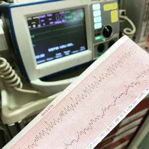
Diagnosis of ACS is time sensitive and done with a thorough history and physical exam, EKG, and cardiac biomarkers. Ideally, the EKG is done within 10 minutes of patient arrival. Even if an EKG is found to be normal, this does not rule out ACS and NSTEMI. The first branch point should be STEMI or not. Typical findings on EKG of an NSTEMI include ST depression, transient ST-elevation, or new T wave inversions. Serial EKG’s at predetermined intervals are common practice. It is also of benefit to repeat the EKG with any new patient symptom or change in the symptoms. It is important to note that even patients with normal or nonspecific ECGs have a 1% to 5% incidence of MI with another 4% to 23% incidence of unstable angina.
“Remember that you don’t need an elevated troponin to diagnosis a STEMI (initial serum markers are sometimes negative).”
Let’s talk the latest gossip…what are the “high sensitivity Troponin” we keep hearing about?
The traditional cardiac troponin was historically quoted to become elevated within 2 to 4 hours after symptom onset. Many facilities are now beginning to measure “high sensitivity troponins”. These allow the ED physician to obtain an initial trop at time 0 and a repeat trop at time 2 hours, instead of the conventional time 0 with the repeat trop at time 3, 6, 12, and 24 hours (depending on hospital practice). The newer high-sensitivity troponins have been shown to identify 90% to 100% of patients with MI at the time of arrival using the lowest cut point. In those that are initially elevated, a delta or changing values over a 1- to 3-hour period can help distinguish acute from chronic injury.
What is the rationale behind this timing?
Routine traditional troponin measurement at 0 and 3 hours in individuals with a HEART score of 3 or less or a TIMI score of 0 can predict a low rate of 30-day MACE (major adverse cardiac event). Low risk correlates to a risk <2% of MACE in the next 30 days. In contrast to the older troponin measurements we are all used to using and mentioned above, one total high sensitivity troponin test below the level of detection at the time of patient arrival OR negative (but detectable) high-sensitivity trop at time 0 hours and 2 hours predicts a low rate of MACE. The European Society of Cardiology recommends a 3-hour serial interval when high-sensitivity troponins are used.
Wait a second, say that again…?
You heard it right; you only need ONE undetectable level with new high sensitivity troponin if the patient has had >3 hours of symptoms. One UNDETECTABLE high-sensitivity troponin at time 0 is enough to predict a low 30-day MACE rate. In addition, a DETECTABLE troponin (but still negative) at time 0 and 2 hours along with a negative “delta” predict a low 30-day MACE rate. In sum, patients with a heart score of 3 or less, a non-ischemic ECG, and negative serial high sensitivity Trop at time 0 and 2 hours have a low MACE rate and can therefore be discharged from the ED.
What about additional testing and follow-up for these low risk patients?
Multiple studies have shown no improvement or benefit in low-risk patients when provocative testing (stress tests, CT angiography) was performed prior to discharge. To address the small percentage of patients that are missed by these guidelines, it is suggested that providers consider recommending patients to follow-up with their primary care physician or cardiologist within 1 to 2 weeks after discharge, even though no studies to date have been able to demonstrate any measurable benefit.
Management:
Back to patients who are high risk for NSTEMI. Unless there are contraindications, NSTEMI patients should be given 324 mg of chewable, non-coated, aspirin as well as heparin or LMWH in the ED. Remember that with aspirin, the estimated number needed to treat is 41 patients, with the number needed to harm (no dangerous bleeding) being 167 patients. The combination of aspirin and heparin is indicated for patients with ACS and currently considered standard of care by the ACA. Recent studies have shed light on the potential minimal benefit from heparin in NSTEMI, and further details can be read here from Rebel EM. Our advice? Follow what your cardiologist wants since they will be deciding on PCI. More than likely, cardiology will want it.
Oxygen is no longer routinely recommended, as it may actually be harmful in a patient without the need for supplemental O2. It is only recommended for patients with an oxygen saturation level <90%, patients with respiratory distress, or those with high-risk features of hypoxemia. Please remove the nasal canula that was prophylactically placed in triage or with EMS.
Patients with continuing symptoms and/or elevated blood pressure can be given 0.4mg of sublingual nitroglycerin every 5 minutes for up to 3 doses or until the patient’s pain is relieved. In addition, some patients may warrant a nitroglycerin drip to stabilize their symptoms and blood pressure. Contraindications to nitro administration include hypotension, recent use of phosphodiesterase inhibitors, and if there is a concern for RV infarction. Policies and protocols vary by the institution regarding the availability of diagnostic angiography and intervention. If available, a cardiology consult and admission to cardiac care units are advised.
Should you also add an anti-platelet agent?
In patients with NSTEMI, giving P2Y12 inhibitors and glycoprotein IIb/IIIa inhibitors before or after cardiac catheterization did not significantly decrease the risk of MACE. It did, however, increase the risk of bleeding. In sum, these antiplatelet agents are less time-sensitive and can be given at the time of cardiac catheterization by the cardiologist instead of in the ED.
References
1. Basit H, Malik A, Huecker MR. Non ST Segment Elevation Myocardial Infarction. [Updated 2020 Oct 15]. In: StatPearls [Internet]. Treasure Island (FL): StatPearls Publishing; 2020 Jan-. Available from: https://www.ncbi.nlm.nih.gov/books/NBK513228/
https://www.aliem.com/2018-acep-policy-non-st-elevation-acs/
2. van ‘t Hof AWJ, Badings E. NSTEMI treatment: should we always follow the guidelines?. Neth Heart J. 2019;27(4):171-175. doi:10.1007/s12471-019-1244-3
3. Chenevier-Gobeaux C, Sebbane M, Meune C, et al. Is high-sensitivity troponin, alone or in combination with copeptin, sensitive enough for ruling out NSTEMI in very early presenters at admission? A post hoc analysis performed in emergency departments. BMJ Open. 2019;9(6):e023994. Published 2019 Jun 16. doi:10.1136/bmjopen-2018-023994
4. Diercks DB, Hollander JE, Chang A. Acute Coronary Syndromes. In: Tintinalli JE, Ma O, Yealy DM, Meckler GD, Stapczynski J, Cline DM, Thomas SH. eds. Tintinalli’s Emergency Medicine: A Comprehensive Study Guide, 9e. McGraw-Hill; Accessed November 01, 2020. https://accessmedicine.mhmedical.com/content.aspx?bookid=2353§ionid=219671081
5. Januzzi JL Jr, Mahler SA, Christenson RH, Rymer J, Newby LK, Body R, Morrow DA, Jaffe AS. Recommendations for Institutions Transitioning to High-Sensitivity Troponin Testing: JACC Scientific Expert Panel. J Am Coll Cardiol. 2019 Mar 12;73(9):1059-1077. doi: 10.1016/j.jacc.2018.12.046. Epub 2019 Feb 21. PMID: 30798981


