Want to experience the greatest in board studying? Check out our interactive question bank podcast- the FIRST of its kind here: emrapidbombs.supercast.com
Authors: Blake Briggs, MD
Peer Reviewer: Mary Claire O’Brien, MD
Objectives: discuss causes of elevated intracranial pressure, presentation, and acute management.
Introduction: Monro-Kellie Doctrine
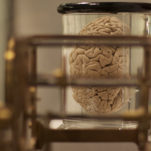
“The sum of the volumes of brain, CSF, and intracranial blood is a constant.” ICP is normally <15 mmHg in adults. Anything >20 mmHg is pathologic. Basic principle: the adult brain is enclosed in a nonexpandable case of bone–the skull. The adult skull has a volume of about 1500 mL, with brain tissue making up about 80% of its contents. The rest is fluid (blood and CSF about 10% each).
If any one of these increases in volume (blood, CSF, or brain tissue), the others must decrease or else ICP will increase (since the cranium only allows for a fixed volume). ICP is normally <15 mmHg in adults. Anything >20 mmHg is pathologic.
As you could imagine, the cranial vault has the ability to be compliant with some increases in ICP via compensatory mechanisms such as CSF displacement into the thecal sac and decrease in blood volume via venoconstriction.
Therefore, the relationship between intracranial volume and ICP is not linear, it’s exponential. Once compensation is exhausted depending on how severe the change in volume to pressure increase is, there will be a profound increase in ICP and a systematic decrease in cerebral blood flow (CBF) and eventual brainstem compression.
Causes: think mass effect. Anything that should not be there inside the cranium will add to intracranial pressure, eventually impairing neurologic function and risking tissue perfusion.
Accelerate your learning with our EM Question Bank Podcast
- Rapid learning
- Interactive questions and answers
- new episodes every week
- Become a valuable supporter
-Trauma brain injury, intracranial bleeding, tumors, hydrocephalus, idiopathic intracranial hypertension, cerebral edema (hepatic encephalopathy, trauma)
CPP = MAP – ICP
Where CPP basically implies CBF. The brainstem exhibits its own level of autoregulation, but this is not possible when there is elevated ICP.
The conditions mentioned above cause an elevated ICP, leading to a corresponding decrease in CPP and eventual brainstem compression and herniation.
Presentation of symptoms: nonspecific headache (if patient is conscious) à CN VI palsies, papilledema à Cushing’s Triad
Cushing’s Triad = increased SBP, decreased pulse, decreased respiratory rate
The actual mechanism of Cushing’s Triad is still debated, but what isn’t debated is how deadly and poorly prognostic it is and what a poor prognosis it indicates. Urgent intervention is indicated.
Management
Goal: ICP monitoring. Placing an intracranial monitor allows an intensivist to maintain CPP and oxygenation of cerebral tissue. The only way to do this is measure MAP (A-line) and ICP (monitor) to derive CPP. The practice of placing these and the desirable anatomical location to place them is beyond the scope of this review.
-ICP monitoring is associated with decreased mortality in traumatic head injury patients.
-Briefly, when is ICP monitoring indicated? GCS <8, concerning presentation for increased ICP, some intracranial process that warrants aggressive care.
In the meantime, there are some maneuvers can assist in acutely, but temporarily, reducing ICP.
Initial resuscitation: These patients should rapidly have their airway secured. Propofol is often used due to fast on/offset.
–Avoid large swings in BP. Hypertension with SBP >200 mmHg is bad but so is a major drop peri-intubation into the SBP <110. Hypotension leads to cranial vasodilation and therefore elevated ICP. Hypotension is known to exacerbate secondary brain injury.
-BP should be maintained for CPP >60 and ICP <20. Only treat HTN if CPP >120 or ICP >20.
Specific BP parameters depending on the cause: (see our handout on hypertensive emergencies for more details)
-Traumatic brain injury: SBP >110
-Intracranial hemorrhage: SBP ~140. If >220, aggressive lowering. Nicardipine, clevidipine, labetalol.
-Subarachnoid hemorrhage: SBP <160 (hotly debated). Same drugs as above. Nimodipine for vasospasm
-Keep patients euvolemic and normal/~hyperosmolar. Normal saline should be used, NO hypotonic fluids. Hyponatremia has poor prognosis.
In addition to Cushing’s Triad, the following clinical signs require urgent intervention:
-GCS <8, bilateral dilated/fixed pupils, decorticate or decerebrate posturing, hypothermia, hypoxia, hypotension.
Immediate things to do:
-Head of bed to 30 degrees or greater if able
-Hyperventilate to PCO2 of 30-35 (effects only last for a few hours so you get one shot at this; the effect is not reproducible if you attempt to do this again in <24 hours and actually can be harmful).
-Glucose: 90-180; tight control is indicated, but hypoglycemia is worse than hyperglycemia
-Reversal of blood thinners:
Warfarin = Vit K and PCC
Dabigatran = Idarucizumab > FEIBA > PCC
Xa inhibitors = Andexanet alfa, PCC
-Temperature: keep afebrile, target 38 Celsius
-Sedation: all patients should be sedated to minimize ventilator asynchrony, metabolic demand, and hypertension.
-Seizure prophylaxis: hotly debated. As of this writing, seizure prophylaxis should be on a case-by-case basis, but there is an increasing consensus to not give it if the patient is not high risk for seizures and has not yet had one in the setting of elevated ICP. The exception is TBI, levetiracetam is preferred for prophylaxis.
-Osmotic diuresis if impending herniation: consider mMannitol or hHypertonic saline
Mannitol 1g/kg. Can be repeated 0.5g/kg every 6-8 hours. Effects occur within minutes and peak in 1 hour. Serial serum osmolality measurements are required.
-Dehydration and renal injury are obvious major issues (especially in trauma).
Hypertonic saline bolus: 3% NaCl bolus, followed by 23.4% NaCl infusion in a central line (due to risk of extravasation). Boluses are preferred over infusions, with 140-150 mmol/L serum Na as the goal.
It has been shown that hypertonic saline has greater efficacy in lowering ICP, but there has been no examination of clinical outcomes. Basically, neither agent has been found to be superior, but hypertonic saline causes less dehydration and renal injury and is easier to dose.
What NOT to use: Furosemide, glucocorticoids (steroids may have a role in vasogenic edema secondary to CNS infections and brain tumors).
More “definitive” therapy: the best intervention for elevated ICP is to actually address the underlying issue. This could include evacuation of a blood clot, mass resection, treatment of an underlying metabolic disorder, Removal of CSF: via ventriculostomy (EVD), or even a decompressive craniectomy (removing bony skull and therefore bypassing the Monro-Kellie doctrine).
References
1. Battison C, Andrews PJ, Graham C, Petty T. Randomized, controlled trial on the effect of a 20% mannitol solution and a 7.5% saline/6% dextran solution on increased intracranial pressure after brain injury. Crit Care Med 2005; 33:196.
2. Francony G, Fauvage B, Falcon D, et al. Equimolar doses of mannitol and hypertonic saline in the treatment of increased intracranial pressure. Crit Care Med 2008; 36:795.
3. Ichai C, Armando G, Orban JC, et al. Sodium lactate versus mannitol in the treatment of intracranial hypertensive episodes in severe traumatic brain-injured patients. Intensive Care Med 2009; 35:471.
4. Kamel H, Navi BB, Nakagawa K, et al. Hypertonic saline versus mannitol for the treatment of elevated intracranial pressure: a meta-analysis of randomized clinical trials. Crit Care Med 2011; 39:554.
5. Mortazavi MM, Romeo AK, Deep A, et al. Hypertonic saline for treating raised intracranial pressure: literature review with meta-analysis. J Neurosurg 2012; 116:210.
6. Schwarz S, Schwab S, Bertram M, et al. Effects of hypertonic saline hydroxyethyl starch solution and mannitol in patients with increased intracranial pressure after stroke. Stroke 1998; 29:1550.
7. Vialet R, Albanèse J, Thomachot L, et al. Isovolume hypertonic solutes (sodium chloride or mannitol) in the treatment of refractory posttraumatic intracranial hypertension: 2 mL/kg 7.5% saline is more effective than 2 mL/kg 20% mannitol. Crit Care Med 2003; 31:1683.


